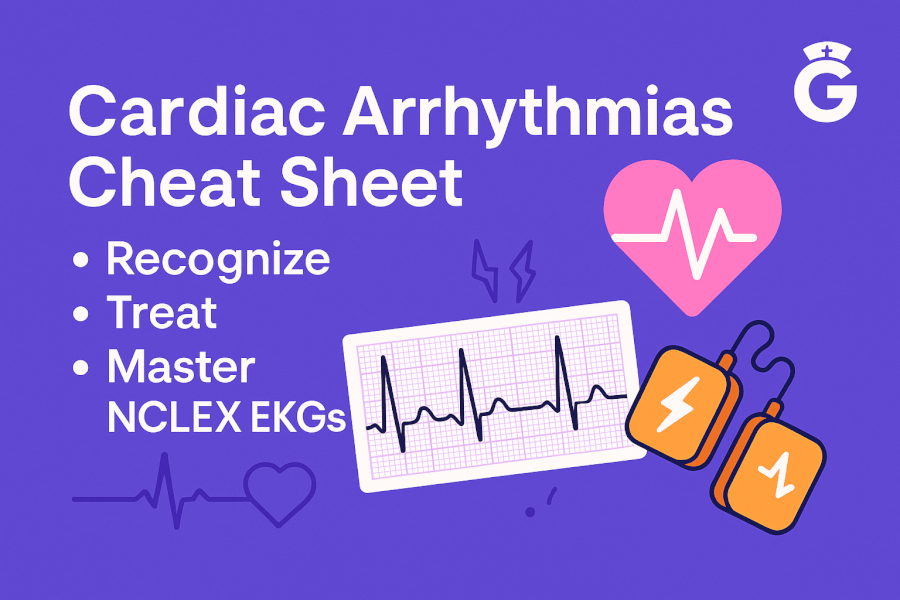

Heart Rhythm Strips Cheat Sheet: Every Cardiac Arrhythmia You Need for the NCLEX
Reading heart rhythm strips is one of the most tested skills on the NCLEX—and one of the most intimidating. This cheat sheet breaks down every cardiac arrhythmia into a scannable, easy-to-review format so you can recognize rhythms fast, recall the right interventions, and walk into your exam with confidence.
Whether you are studying EKG strips for the first time or reviewing before test day, bookmark this page. We cover normal sinus rhythm through lethal arrhythmias, with a quick-reference table, mnemonics, and nursing priorities for each rhythm.
🎯 Free NCLEX quiz!
See how well you can identify key cardiac rhythms.
Quick-Reference: Heart Rhythm Strips at a Glance
Use this table as your go-to study aid. Each row summarizes the rhythm, rate, key EKG features, and the first-line intervention.
| Rhythm | Rate (bpm) | Regularity | P-Waves | QRS Width | Priority Intervention |
|---|---|---|---|---|---|
| Normal Sinus Rhythm | 60–100 | Regular | Present, upright | Narrow | None needed |
| Sinus Bradycardia | < 60 | Regular | Present, upright | Narrow | Atropine if symptomatic |
| Sinus Tachycardia | > 100 | Regular | Present, upright | Narrow | Treat underlying cause |
| Atrial Fibrillation | Varies (atrial 300–600) | Irregularly irregular | Absent / fibrillatory | Narrow | Rate control + anticoagulation |
| Atrial Flutter | Atrial 250–350 | Regular or regularly irregular | Sawtooth flutter waves | Narrow | Rate control + anticoagulation |
| SVT | 150–250 | Regular | Often hidden in T-wave | Narrow | Vagal maneuvers → adenosine |
| Junctional Escape | 40–60 | Regular | Absent, inverted, or after QRS | Narrow | Atropine or pacing if symptomatic |
| PVCs | Underlying rate | Irregular (early beats) | Absent before PVC | Wide | Correct K⁺/Mg²⁺; amiodarone if frequent |
| Ventricular Tachycardia | 100–250 | Regular | Usually absent | Wide | With pulse: amiodarone or cardioversion. Pulseless: CPR + defib |
| Ventricular Fibrillation | None (chaotic) | Chaotic | None | No true QRS | Immediate CPR + defibrillation |
| Asystole | None (flatline) | None | None | None | CPR + epinephrine (do NOT shock) |
| 1st-Degree AV Block | 60–100 | Regular | Present, prolonged PR > 0.20 s | Narrow | Monitor; treat cause |
| 2nd-Degree Type I (Wenckebach) | Usually < 100 | Irregular (progressive) | PR lengthens then drops | Narrow | Monitor; atropine if symptomatic |
| 2nd-Degree Type II (Mobitz II) | Usually < 100 | Irregular (sudden drop) | Fixed PR, dropped QRS | Narrow or wide | Pacing (can progress to 3rd-degree) |
| 3rd-Degree (Complete) AV Block | Usually 20–60 | Regular but dissociated | P-waves march through independently | Wide | Immediate pacing |
Tip: Print or screenshot this table for clinical rotations. Having a quick visual reference while you practice reading rhythm strips makes a huge difference.
The Cardiac Conduction System: A 60-Second Refresher
Before you can read heart rhythm strips, you need to know how the electrical signal normally travels through the heart. Here is the pathway in order:
- SA node (sinoatrial node) in the right atrium fires the impulse—this is the heart’s natural pacemaker.
- The impulse spreads across both atria, producing the P-wave on the EKG strip.
- AV node (atrioventricular node) receives the signal and briefly delays it so the ventricles can fill with blood. This delay is the PR interval (normal: 0.12–0.20 seconds).
- The impulse shoots down the Bundle of His and splits into the right and left bundle branches.
- Purkinje fibers distribute the signal through the ventricles, causing ventricular contraction—this produces the QRS complex (normal: < 0.12 seconds).
- The ventricles repolarize, creating the T-wave.
When any part of this pathway malfunctions, you get an arrhythmia. Knowing where the problem originates tells you which rhythm you are looking at and which intervention to choose.
Sinus Rhythms
Normal Sinus Rhythm (NSR)
- Rate: 60–100 bpm
- Rhythm: Regular
- P-waves: One upright P-wave before every QRS
- PR interval: 0.12–0.20 seconds
- QRS: Narrow (< 0.12 s)
- Intervention: None—this is the normal rhythm
NSR is your baseline. Every other rhythm on this cheat sheet is a deviation from this pattern, so know it cold.
Sinus Bradycardia
- Rate: < 60 bpm
- Strip appearance: Looks like NSR but slower
- Common causes: Athletic heart, beta-blockers, calcium channel blockers, hypothyroidism, vagal stimulation
- Intervention: If the patient is symptomatic (hypotension, dizziness, altered LOC), give atropine 0.5 mg IV. If atropine fails, consider transcutaneous pacing.
NCLEX pearl: The question will test whether you treat the number or the patient. A well-conditioned athlete with a heart rate of 52 who is asymptomatic does not need atropine.
Sinus Tachycardia
- Rate: > 100 bpm (usually 100–160)
- Strip appearance: Looks like NSR but faster
- Common causes: Fever, pain, anxiety, hypovolemia, anemia, infection, hyperthyroidism
- Intervention: Treat the underlying cause. Sinus tach is a symptom, not a primary problem.
Sinus Arrhythmia
- Rate: 60–100 bpm
- Rhythm: Slightly irregular, increases with inspiration, decreases with expiration
- Intervention: None—this is a normal finding, especially in younger patients
Atrial Rhythms
Atrial Fibrillation (A-Fib)
- Rate: Atrial 300–600 (chaotic); ventricular varies
- Rhythm: Irregularly irregular (the hallmark finding)
- P-waves: None—replaced by chaotic fibrillatory waves
- QRS: Narrow (unless aberrant conduction)
- Key risks: Loss of atrial kick reduces cardiac output by up to 25%; blood pools in the atria, forming clots → stroke risk
- Interventions:
- Rate control: Beta-blockers (metoprolol), calcium channel blockers (diltiazem), or digoxin
- Rhythm control: Amiodarone, cardioversion
- Anticoagulation: Warfarin, apixaban, rivaroxaban to prevent stroke
- Assess for signs of hemodynamic instability
Mnemonic: "A-fib is a quivering jelly"—the atria quiver instead of contracting effectively. The rhythm on the strip looks chaotic with no repeating pattern.
Atrial Flutter
- Rate: Atrial 250–350 bpm; ventricular depends on AV conduction ratio (commonly 2:1, so ventricular rate ~150)
- Rhythm: Regular or regularly irregular
- P-waves: Classic sawtooth pattern (flutter waves)
- QRS: Narrow
- Interventions: Similar to A-fib—rate control, anticoagulation, possible ablation
Mnemonic: "Flutter has a sawtooth pattern"—if you see those characteristic zigzag waves between QRS complexes, it is flutter.
Supraventricular Tachycardia (SVT)
- Rate: 150–250 bpm
- Rhythm: Regular with narrow QRS
- P-waves: Often buried in the preceding T-wave and not visible
- Symptoms: Sudden onset palpitations, dizziness, chest tightness
- Interventions (in order):
- Vagal maneuvers (bearing down, carotid massage, cold water to the face)
- Adenosine 6 mg rapid IV push (follow with 12 mg if no conversion)
- Beta-blockers or calcium channel blockers
- Synchronized cardioversion if hemodynamically unstable
Mnemonic: "SVT → Stop Via Tricks (vagal), then Adenosine"
Junctional Rhythms
When the SA node fails or slows, the AV junction can take over as a backup pacemaker. Junctional rhythms have a rate of 40–60 bpm and typically show absent, inverted, or retrograde P-waves.
Premature Junctional Contractions (PJCs)
- Appearance: Early beat with inverted or absent P-wave; narrow QRS
- Cause: Caffeine, stress, digoxin toxicity
- Intervention: Usually benign; treat the underlying cause
Junctional Escape Rhythm
- Rate: 40–60 bpm
- P-waves: Absent, inverted before QRS, or seen after QRS
- Intervention: Atropine or pacing if symptomatic
Accelerated Junctional Rhythm
- Rate: 60–100 bpm (faster than expected for junctional)
- Intervention: Usually monitored; treat if symptomatic
Ventricular Rhythms
Ventricular rhythms originate below the AV node and produce wide QRS complexes (≥ 0.12 seconds). They range from benign (occasional PVCs) to immediately lethal (V-fib).
Key rule for heart rhythm strips: Wide QRS = think ventricular.
Premature Ventricular Contractions (PVCs)
- Appearance: Early, wide, bizarre QRS complex with no preceding P-wave; followed by a compensatory pause
- Common causes: Hypoxia, hypokalemia, hypomagnesemia, caffeine, anxiety
- When to worry: PVCs that are frequent (> 6/min), multifocal, occur in runs of 3+, or fall on the T-wave ("R-on-T phenomenon")
- Intervention: Correct electrolytes (K⁺ and Mg²⁺ first); if persistent and symptomatic, amiodarone
Ventricular Tachycardia (V-Tach)
- Rate: 100–250 bpm
- Rhythm: Usually regular
- QRS: Wide (≥ 0.12 s); may be monomorphic or polymorphic
- Two critical scenarios:
- With pulse (stable): Amiodarone IV or synchronized cardioversion
- Pulseless: Treat as cardiac arrest → CPR + defibrillation + epinephrine + amiodarone per ACLS
- Special case—Torsades de Pointes: A polymorphic VT associated with prolonged QT interval. First-line treatment is IV magnesium sulfate 2 g.
Ventricular Fibrillation (V-Fib)
- Strip appearance: Chaotic, disorganized waves—no identifiable P-waves, QRS, or T-waves
- Cardiac output: NONE—this is a lethal rhythm
- Intervention: Immediate CPR and defibrillation. Follow ACLS: epinephrine every 3–5 minutes, amiodarone after third shock.
NCLEX pearl: V-fib and pulseless V-tach are the only two shockable rhythms. Asystole and PEA are NOT shockable.
Asystole
- Strip appearance: Flat line (confirm in two leads to rule out lead disconnect)
- Cardiac output: NONE
- Intervention: High-quality CPR, epinephrine every 3–5 minutes. Do NOT defibrillate—there is no electrical activity to reset.
Heart Blocks: First Through Third Degree
Heart blocks occur when conduction between the atria and ventricles is delayed or completely interrupted at the AV node or below.
First-Degree AV Block
- PR interval: Prolonged (> 0.20 s) but constant
- Every P-wave is followed by a QRS
- Causes: Digoxin, beta-blockers, aging, ischemia
- Intervention: Monitor; usually benign. Treat the cause if medication-related.
Second-Degree AV Block Type I (Mobitz I / Wenckebach)
- Pattern: PR interval gets progressively longer until a QRS is dropped, then the cycle repeats
- Mnemonic: "Longer, longer, longer, DROP—that's a Wenckebach!"
- Intervention: Usually stable; atropine if symptomatic
Second-Degree AV Block Type II (Mobitz II)
- Pattern: PR interval is fixed and constant, but QRS complexes are randomly dropped
- Why it matters: Much more dangerous than Type I—can suddenly progress to third-degree block
- Intervention: Prepare for pacing. This patient needs a pacemaker.
NCLEX tip: Mobitz II is the "sneaky" block. The PR looks normal, then the QRS just disappears without warning.
Third-Degree (Complete) AV Block
- Pattern: P-waves and QRS complexes march at their own rates with no relationship to each other (AV dissociation)
- Ventricular rate: Usually very slow (20–40 bpm if escape rhythm is ventricular)
- QRS: Often wide (ventricular escape) but can be narrow (junctional escape)
- Symptoms: Severe bradycardia, syncope, hypotension, altered LOC
- Intervention: Immediate transcutaneous pacing followed by permanent pacemaker placement
Nursing Priorities When You Identify an Abnormal Rhythm
No matter which arrhythmia you identify on a heart rhythm strip, your nursing response follows the same decision framework:
- Assess the patient first, not just the monitor. Is the patient symptomatic? Check level of consciousness, blood pressure, SpO₂, and whether they report chest pain or dizziness.
- ABCs always come first. For any unstable patient—secure the airway, support breathing, and address circulation.
- Know your shockable vs. non-shockable rhythms. V-fib and pulseless V-tach → defibrillate. Asystole and PEA → CPR + epinephrine only.
- Correct reversible causes (the H’s and T’s): Hypovolemia, hypoxia, hydrogen ion (acidosis), hypo/hyperkalemia, hypothermia, tension pneumothorax, tamponade, toxins, thrombosis (pulmonary and coronary).
- Document: Note the rhythm, rate, patient symptoms, interventions given, and patient response.
Mnemonics to Remember Heart Rhythm Strips
- "Irregularly irregular = A-fib." If the rhythm has no pattern at all, think atrial fibrillation first.
- "Sawtooth = flutter." Those repeating zigzag waves between QRS complexes mean atrial flutter.
- "Wide QRS = ventricular." Any rhythm with a wide QRS complex (≥ 0.12 s) likely originates in the ventricles.
- "Longer, longer, longer, DROP = Wenckebach." The PR interval progressively lengthens until a beat is dropped.
- "No P’s, narrow QRS = junctional." If P-waves are missing or inverted but the QRS is narrow, the rhythm comes from the AV junction.
- "Shock V-fib and pulseless V-tach; don’t shock asystole." Only defibrillate rhythms that have disorganized electrical activity.
- "SVT → Stop Via Tricks, then Adenosine." Try vagal maneuvers first for SVT before pushing adenosine.
NCLEX-Style Practice: Test Your Rhythm Recognition
Question 1: A patient on the telemetry unit has a heart rate of 42 bpm with a regular rhythm and normal P-waves preceding each QRS. The patient reports feeling lightheaded. What is your priority action?
Answer: This is symptomatic sinus bradycardia. Administer atropine 0.5 mg IV as the first-line intervention, and prepare for transcutaneous pacing if atropine is ineffective.
Question 2: You are monitoring a post-MI patient who suddenly shows a wide-complex tachycardia at 180 bpm with a pulse. Blood pressure is 88/60 mmHg. What do you do first?
Answer: This is ventricular tachycardia with a pulse and hemodynamic instability. Prepare for synchronized cardioversion immediately. Amiodarone can be given if the patient is more stable.
Question 3: A patient’s rhythm strip shows no identifiable P-waves, an irregularly irregular ventricular rhythm at 110 bpm, and narrow QRS complexes. What rhythm is this?
Answer: Atrial fibrillation with rapid ventricular response. The hallmark is the irregularly irregular rhythm with absent P-waves. Priority: rate control (diltiazem or metoprolol) and assess for anticoagulation needs.
Want more practice? Try our full NCLEX practice quiz with detailed rationales →
Related GoodNurse Resources
Build on your heart rhythm strip knowledge with these companion guides:
- Pharmacology Mnemonics: A Complete Guide — Master cardiac drugs including beta-blockers, calcium channel blockers, and antiarrhythmics
- Electrolyte Imbalance Cheat Sheet — Electrolyte imbalances are the #1 cause of arrhythmias; know them cold
- Normal Lab Values Cheat Sheet for Nursing Students — Quick reference for potassium, magnesium, calcium, and other values that affect heart rhythms
- Fluid and Electrolyte NCLEX Questions — Practice questions on the electrolyte disorders that cause arrhythmias
- 50 NCLEX Questions With Answers and Detailed Explanations — Mixed-difficulty practice including cardiac scenarios
By learning to read heart rhythm strips systematically—checking rate, regularity, P-waves, PR interval, and QRS width in that order—you will be able to identify any arrhythmia the NCLEX throws at you. Keep practicing with real rhythm strips, review this cheat sheet before your exam, and use our free NCLEX practice questions to test yourself under timed conditions.
2,500+ NCLEX-style questions with detailed rationales, adaptive difficulty, and AI-powered explanations that break down why each answer is right or wrong. Available in 10+ languages.
Under $10/month — no card needed to start.
Start Free Today →
Download our free printable ECG/EKG Interpretation cheat sheet. Enter your email below and we'll send it right over.