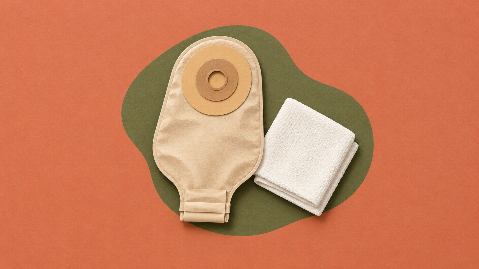


A colostomy brings part of the colon to an abdominal opening to pass stool. An ileostomy brings the ileum, the last part of the small intestine, to that opening to pass intestinal contents. A urostomy drains urine through an abdominal opening. The quickest way to distinguish them is to identify the pathway and what leaves it, rather than judge the pouch from its appearance. NIDDK: bowel ostomy types, NIDDK: urinary diversion.
For nursing students, a particularly confusing example is the ileal conduit: it uses a segment of small intestine to carry urine, but it is a urinary diversion, not an ileostomy carrying stool. That difference changes how you interpret the output.
This is a concept review for learning and supervised clinical discussion. A person's operative record, care plan, and ostomy team determine their actual anatomy and care.
Colostomy vs ileostomy vs urostomy at a glance
| Type | Pathway to remember | Output to expect | Key learning question |
|---|---|---|---|
| Colostomy | Colon to abdominal stoma | Stool; consistency varies with the remaining bowel | Which part of the colon is involved? |
| Ileostomy | Ileum to abdominal stoma | Intestinal contents, typically looser than colostomy stool | How does bypassing the colon affect water absorption? |
| Urostomy | Urinary tract to abdominal stoma | Urine | What urinary diversion was created? |
These are broad patterns, not instructions for diagnosing an unfamiliar stoma. The United Ostomy Associations of America's terminology guide distinguishes stool-draining ostomies from urine-draining ostomies and emphasizes describing the output clearly.
Start with the vocabulary: ostomy, stoma, and pouch
Ostomy refers to the surgically created opening; stoma names the opening itself. The pouching system collects output outside the body. Its skin barrier helps protect the surrounding skin from contact with the output. One-piece and two-piece systems exist, so the number of visible components does not tell you which organ is connected. NIDDK: managing bowel ostomy output.
Keep three separate lines in your notes:
- Anatomy: What structure connects to the stoma?
- Output: What is leaving, and what has changed?
- Appliance: What system is collecting it?
This separation helps prevent a common reasoning error: seeing a familiar-looking bag and assuming a familiar internal route.
Colostomy: the colon is the clue
A colostomy involves the large bowel. The NHS explains that a segment of colon is brought through the abdominal wall to form a stoma. It may be temporary or permanent; the name alone does not establish whether it will be reversed. NHS: how a colostomy is done.
Stool consistency is not identical in every colostomy. The colon normally absorbs water, so how much bowel remains in the pathway matters. Avoid memorizing “all colostomies produce solid stool” as a rule. NIDDK: digestive changes after surgery.
For a classroom report, “colostomy” is a starting point. A stronger report adds the documented location, what the output actually looks like, and whether that differs from the person's usual pattern. Do not fill missing operative details with a guess based on which side of the abdomen holds the pouch.
Ileostomy: connect the route to fluid balance
An ileostomy brings the ileum through the abdominal wall. In this pathway, the colon is bypassed or removed. This is why the word ileum deserves more attention than the appearance of the appliance. NIDDK: ileostomy anatomy.
Output is generally liquid to pasty rather than reliably formed. Bypassing the colon reduces the opportunity for water absorption, and dehydration is a recognized complication of bowel ostomies, especially when the large intestine is removed or bypassed. UOAA: output descriptions, NIDDK: ostomy complications.
The learning point is to connect anatomy with assessment. “There is output” is less informative than a description of its amount, consistency, trend, and associated symptoms. A student should discuss concerning changes with the supervising nurse and follow the individualized plan rather than apply a universal fluid prescription or output cutoff from a study card.
Urostomy: why an ileal conduit still drains urine
In an ileal conduit, surgeons use a separated segment of intestine as a passageway. Urine travels from the ureters through that segment and exits through the stoma into an external pouch. The segment serves the urinary route; it is not carrying food through the digestive tract. NIDDK: urostomy and ileal conduit.
That makes “it contains ileum, therefore it must be an ileostomy” incorrect reasoning. Ask what travels through the segment.
Not every urinary diversion has the same construction or emptying method. NIDDK also describes cutaneous ureterostomy and continent urinary diversions. Learn the documented procedure before transferring assumptions from one type to another. For another introduction to the urinary purpose of these openings, see the American Cancer Society's urostomy overview.
Nursing observations: describe the change before naming a cause
A focused learning checklist can organize what to communicate:
- Confirm the documented type. Separate the surgery name from what you can observe.
- Describe the stoma and surrounding skin. Keep the stoma itself distinct from skin irritation or leakage around the appliance.
- Describe output over time. Include the observed amount and consistency, with the time interval used.
- Ask about associated symptoms and concerns. Use the person's own description without assuming the cause.
- Identify what needs prompt escalation. Communicate new or concerning findings to the supervising nurse and ostomy team.
Skin irritation, impaired blood supply, dehydration, and bowel obstruction are among recognized bowel-ostomy complications. Heavy or persistent bleeding, severe vomiting, or absent stool/gas with abdominal symptoms warrant urgent clinical assessment. These examples are not a complete triage list or permission to wait for a particular threshold. NIDDK: complications and warning signs.
The UOAA guide supports using specific descriptions of the stoma, skin, and output. For help structuring those observations into a report, review GoodNurse's SBAR practice guide.
Three original practice checks
These fictional learning examples are not official NCLEX questions.
1. The record says “ileal conduit.” Is the pouch collecting stool?
No. The key information is the documented urinary diversion. Explain the route of urine before considering what the pouch looks like.
2. A student says every colostomy produces formed stool. What is missing?
The location and remaining bowel matter. A broad category label should not replace observation of the actual output or knowledge of the documented anatomy.
3. You see an abdominal pouch, but the surgery name is unavailable. Can you identify the type confidently?
No. Record what is visible, identify the missing information, and verify the procedure with the supervising nurse or record. An honest unknown is more useful than an unsupported label.
Use these checks to explain the route aloud: colon and stool; ileum and intestinal contents; urinary diversion and urine. Then add the person's documented details. For a related elimination concept, read urinary retention vs incontinence.
AI-assistance note: This article was prepared with AI assistance using the linked public sources. The illustrative cover is AI-generated. No human clinical review is claimed.