

An arterial leg ulcer develops when too little blood reaches the tissue. A venous leg ulcer develops when blood has trouble returning through the leg veins and pressure builds in them. A sore on a cool toe points you toward arterial flow; a shallow, draining sore near a swollen inner ankle points you toward venous disease. Both patterns can occur in the same leg, so the wound’s appearance alone cannot tell you whether compression is safe. See the Society for Vascular Surgery’s guidance on venous disease and the Wound, Ostomy, and Continence Nurses Society’s guidance.
Start with the direction of blood flow
Arterial: too little gets in. Arteries supply the skin and deeper tissues with oxygenated blood. When peripheral artery disease (PAD) narrows that route, even a small injury can be slow to heal. Toes and feet are common places to notice a nonhealing sore. The patient might describe calf discomfort that comes on while walking and settles with rest, or foot pain even at rest in more advanced disease. A foot that is colder than the other adds to the concern. NHLBI’s guide to PAD symptoms explains why some people have atypical symptoms or no typical warning pattern.
Venous: blood has trouble getting back. In the legs, one-way valves and the calf muscles help move blood toward the heart. When valves leak or a vein is obstructed, pressure in the veins can stay high. Over time, swelling and changes in the surrounding skin can accompany breakdown. This helps explain why a wound just above the inner ankle, with swelling and brown discoloration around it, raises a different question from a sore on a cool toe. See the Society for Vascular Surgery’s guide to lower-extremity venous disease.
Compare the wound and the rest of the leg
These are common patterns to organize your assessment. No single finding confirms the cause. Compare them with NHLBI’s description of PAD symptoms and the American Academy of Family Physicians’ comparison of chronic wounds.
| Assessment | Arterial clues | Venous clues |
|---|---|---|
| Wound location | Toes or foot; may also occur around the ankle | Lower leg, often just above the inner ankle |
| Wound appearance | May have sharply defined edges and a dry or dark base | Often shallow and draining, with irregular edges |
| Surrounding leg and foot | Cooler foot or weaker foot (pedal) pulses | Ankle swelling, brown discoloration, or thickened skin |
| History | Leg discomfort with walking or foot pain at rest | Longstanding swelling, varicose veins, or prior deep vein thrombosis |
Describe what you see before naming what it might mean. “Shallow wound above the right inner ankle with drainage and surrounding swelling” gives the next clinician more information than “looks venous.” In a supervised assessment, note when the wound started, how it has changed, its location and size, the tissue and drainage you can see, and what you found when you compared the feet. If you couldn’t feel a pulse, report where you checked and that it wasn’t palpable on your exam. That finding deserves follow-up; it doesn’t prove the artery is blocked.
Why the team checks arterial flow before compression
Compression can support healing of a venous ulcer, but significant arterial insufficiency changes what is safe. Swelling or a wound in a typical venous location does not rule out poor arterial flow. The WOCN compression algorithm accounts for arterial findings, including when mixed disease requires a different plan. In clinical placement, raise conflicting findings with your supervising nurse and confirm the ordered plan before applying compression.
One common test is the ankle-brachial index (ABI), which compares systolic blood pressure at the ankle with systolic pressure in the arm. A low result can support concern about reduced arterial flow. An unexpectedly high result isn’t automatically reassuring: in some people with diabetes or chronic kidney disease, stiff arteries can resist compression by the cuff and make the reading falsely high. The team may use toe pressures or a toe-brachial index when the ankle arteries are noncompressible. NHLBI’s overview of PAD diagnosis describes both tests.
Practice with three fictional findings
- Swollen ankle, draining wound, cool foot. The location and swelling suggest a venous component. The cool foot adds an arterial concern. In your report, include both and ask your supervising nurse whether arterial perfusion has been assessed before compression.
- A toe wound that hasn’t healed, with calf pain during walks that eases with rest. Together, these clues raise concern about PAD. Report the walking pattern and what you found on the foot assessment. A wound on a toe alone could have other explanations.
- A foot becomes suddenly cold, painful, and numb. The change from baseline matters more than classifying the old wound. This could be acute limb ischemia, a vascular emergency. Escalate immediately through your clinical team’s emergency process rather than waiting for routine wound rounds. See the Society for Vascular Surgery’s guide to acute limb ischemia.
To rehearse the first scenario, use the GoodNurse SBAR practice guide. Try stating the wound findings, the reason you’re concerned about flow, and the specific information you need before compression. If terms like necrosis or pallor are unfamiliar, see the GoodNurse nursing vocabulary guide.
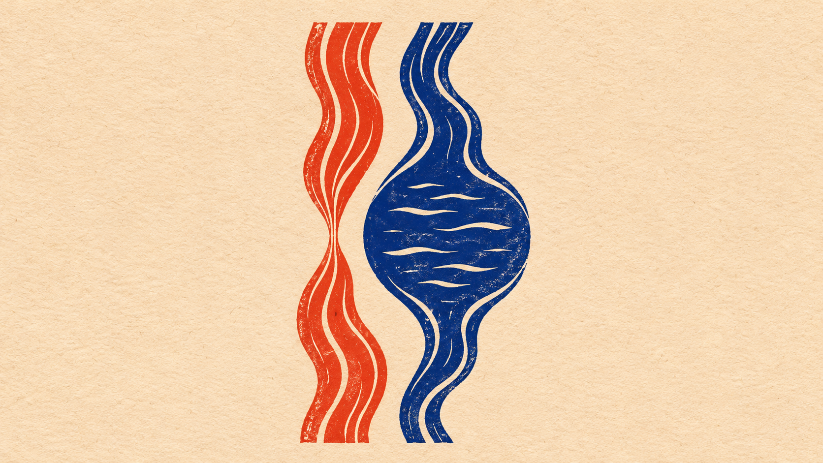
AI assisted with drafting this nursing education article and generated the cover illustration.