Preparing for the NCLEX exam can be a daunting task, especially when it comes to mastering complex topics like Electrocardiograms (ECGs). Understanding ECGs is crucial for any aspiring nurse, as it plays a vital role in diagnosing and managing cardiac conditions. This comprehensive guide will walk you through the essentials of ECG interpretation, and as a bonus, we'll provide you with a handy mnemonic to make learning easier!
💡 Ultimate NCLEX Study Mega Guide
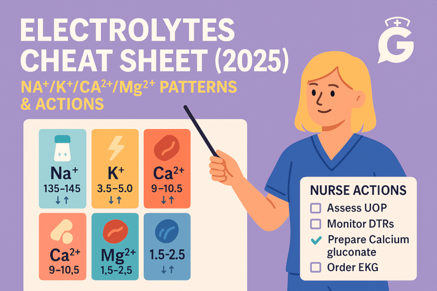
Your must‑have resource to NCLEX cheat sheets, mnemonics & key topics - all in one place.
What is an Electrocardiogram (ECG)?
An Electrocardiogram (ECG or EKG) is a non-invasive test that records the electrical activity of the heart over a period of time. It is a crucial tool in diagnosing various cardiac conditions such as arrhythmias, myocardial infarctions, and electrolyte imbalances. The ECG provides a visual representation of the heart's electrical activity, which can be analyzed to detect abnormalities.
Understanding the Basics of ECG
The Cardiac Cycle
The cardiac cycle consists of a series of electrical events that occur during each heartbeat. These events are represented on the ECG as waves and intervals:
- P Wave: Represents atrial depolarization.
- QRS Complex: Represents ventricular depolarization.
- T Wave: Represents ventricular repolarization.
- U Wave: Sometimes seen after the T wave, represents repolarization of the Purkinje fibers.
ECG Leads
ECG leads are electrodes placed on the body to measure the heart's electrical activity from different angles. There are 12 standard leads:
- Limb Leads: I, II, III, aVR, aVL, aVF
- Precordial Leads: V1, V2, V3, V4, V5, V6
ECG Paper
ECG paper is divided into small and large squares. Each small square represents 0.04 seconds, and each large square (comprising five small squares) represents 0.20 seconds. This grid helps in measuring the duration and intervals of the waves.
Steps to Interpret an ECG
Step 1: Check the Heart Rate
The heart rate can be calculated by counting the number of QRS complexes in a 6-second strip and multiplying by 10. Alternatively, the "300 rule" can be used: divide 300 by the number of large squares between two consecutive R waves.
Step 2: Assess the Rhythm
Determine if the rhythm is regular or irregular by measuring the intervals between R waves. A regular rhythm will have consistent intervals, while an irregular rhythm will have varying intervals.
Step 3: Analyze the P Waves
Check if P waves are present and if they occur before each QRS complex. This helps in identifying atrial activity and determining if the rhythm is sinus.
Step 4: Measure the PR Interval
The PR interval represents the time taken for the electrical impulse to travel from the atria to the ventricles. It should be between 0.12 and 0.20 seconds (3-5 small squares).
Step 5: Evaluate the QRS Complex
The QRS complex duration should be between 0.06 and 0.10 seconds (1.5-2.5 small squares). A wider QRS complex may indicate a ventricular conduction delay.
Step 6: Examine the ST Segment and T Wave
The ST segment should be flat and isoelectric. Elevation or depression of the ST segment can indicate myocardial ischemia or infarction. The T wave should be upright in most leads; inverted T waves can suggest ischemia.
Step 7: Look for Abnormalities
Identify any abnormal waves or patterns, such as Q waves, which may indicate a previous myocardial infarction, or U waves, which can be associated with hypokalemia.
Common ECG Abnormalities
Sinus Bradycardia
- Heart Rate: < 60 bpm
- Rhythm: Regular
- P Wave: Present before each QRS complex
- PR Interval: Normal
- QRS Complex: Normal
Sinus Tachycardia
- Heart Rate: > 100 bpm
- Rhythm: Regular
- P Wave: Present before each QRS complex
- PR Interval: Normal
- QRS Complex: Normal
Atrial Fibrillation
- Heart Rate: Variable
- Rhythm: Irregularly irregular
- P Wave: Absent, replaced by fibrillatory waves
- PR Interval: Not measurable
- QRS Complex: Normal
Ventricular Tachycardia
- Heart Rate: > 100 bpm
- Rhythm: Regular
- P Wave: Absent or not related to QRS complex
- PR Interval: Not measurable
- QRS Complex: Wide (> 0.12 seconds)
Myocardial Infarction
- ST Segment: Elevation or depression
- Q Wave: Pathological Q waves may be present
- T Wave: Inversion
Bonus ECG Mnemonic: "PQRST"
To help you remember the key components of ECG interpretation, use the mnemonic "PQRST":
- P: P wave (Atrial depolarization)
- Q: QRS complex (Ventricular depolarization)
- R: Rhythm (Regular or irregular)
- S: ST segment (Elevation or depression)
- T: T wave (Ventricular repolarization)
Conclusion
Mastering ECG interpretation is essential for any nursing professional, and it can significantly impact patient care. By following the steps outlined in this guide and using the "PQRST" mnemonic, you'll be well-prepared to tackle ECG questions on the NCLEX exam. Remember, practice makes perfect, so keep practicing ECG interpretation to build your confidence and proficiency.
Good luck with your NCLEX preparation, and happy studying!
Download our free printable ECG/EKG Interpretation cheat sheet. Enter your email below and we'll send it right over.