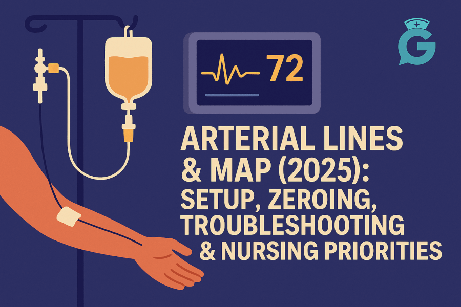
Arterial lines (a-lines) give second-to-second blood pressure and easy access for labs—but only if the setup is correct and the waveform is trustworthy. This guide shows you how to set up and zero, recognize good vs artifact waveforms, pick first actions for common problems, and select parameters that prove your action worked—exactly how NGN expects you to think.
If acid–base and perfusion are part of the picture, keep ABG Interpretation Made Simple (2026) open. For shock trending and organ cross-talk, pair with Lactate in Sepsis (2026) and Cardiac Markers (2026): Troponin & BNP. When a case stem overloads you, reset with How to Read NGN Case Stems (2026).
Table of Contents
- What an Arterial Line Measures (and Doesn’t)
- MAP Targets & Why They Matter
- Setup: Flush System, Leveling, Zeroing
- Waveform Quality: Recognize Good vs Artifact
- Common Problems & First Actions
- Sampling Blood Safely
- NGN Tie-Ins: Action → Parameter Pairs
- Mini Practice Cases (with Answers)
- FAQs
- Further Reading
🎯 Free NCLEX quiz!
Test your knowledge - new quizzes added weekly!
What an Arterial Line Measures (and Doesn’t)
Measures: Beat-to-beat arterial pressure and waveform (systolic/diastolic, MAP), enabling real-time trend and frequent sampling (ABG, lactate).
Doesn’t measure: Central venous pressure, preload, or cardiac output directly—interpret with clinical picture, labs, and echo/hemodynamics as available.
Professor’s note: Don’t let a pretty number fool you. A misleveled transducer can show a “normal” MAP while organs under-perfuse. Trust waveforms and technique.
MAP Targets & Why They Matter
- MAP (mean arterial pressure) reflects organ perfusion better than the systolic alone.
- Common ICU target is MAP ≥ 65 mmHg, but goals can vary (renal disease, neuro cases). Always follow team targets and local protocols.
- Pair MAP with urine output (UOP), mentation, skin temperature, and lactate trend to confirm that perfusion is actually improving.
For organ markers that move with perfusion changes, see Renal Function & UA Interpretation (2026) and Coagulation Studies (2026).
Setup: Flush System, Leveling, Zeroing
1) Flush system
- Pressurized bag at 300 mmHg, continuous flush delivering ~3 mL/hr.
- Eliminate air; tighten connections; secure tubing to avoid traction on the catheter.
2) Level the transducer
- Reference point: phlebostatic axis (4th intercostal space, mid-axillary line).
- Level any time the bed height changes or client position changes significantly.
3) Zero the transducer
- Turn stopcock off to the patient and open to air; press zero on the monitor; close to air and reopen to patient.
- Re-zero if you suspect drift or after component changes.
4) Perform a square-wave test (fast flush)
- Briefly activate the fast-flush device.
- Look for 1–2 oscillations after the square wave:
- Over-damped (sluggish, low systolic/false low pulse pressure) → often due to air, clots, kinks.
- Under-damped (exaggerated peaks, overshoot) → often due to too-stiff tubing, long tubing, or loose connections.
Waveform Quality: Recognize Good vs Artifact
- Normal a-line waveform: rapid upstroke (systole), dicrotic notch (aortic valve closure), downstroke (diastole).
- Over-damped: low systolic, high diastolic, notch blunted—fix the system (air, clot, kink, filter).
- Under-damped: falsely high systolic with ringing—shorten/secure tubing, check connections.
Tip: Always correlate with a manual cuff if numbers don’t fit the clinical picture.
Common Problems & First Actions
-
MAP reads low, client looks okay
Actions: verify level/zero, check square-wave test, compare with cuff; assess perfusion (UOP, mentation, cap refill).
Parameters: MAP (post-fix), UOP, mentation. -
Dampened waveform after line repositioning
Actions: inspect for air/kinks, flush per policy, check dressing/splint; re-zero and re-level.
Parameters: waveform morphology, MAP, SpO₂, symptoms. -
Bleeding at insertion site
Actions: reinforce dressing, check line security, apply gentle pressure, notify provider if persistent; evaluate coagulation status (see Coagulation Studies).
Parameters: visible bleeding, MAP/HR, Hgb trend as ordered. -
Suspected infection (erythema, tenderness, fever)
Actions: notify provider; follow policy for culture/line change; maintain strict asepsis during access.
Parameters: temperature, WBC trend, local site checks. -
Accidental disconnection
Actions: clamp/secure immediately; maintain sterility; monitor hemodynamics; replace per policy.
Parameters: MAP/HR, symptoms, insertion site integrity.
Sampling Blood Safely
- Pause infusions running through the sampling port if required by policy; use waste volume per protocol before the actual sample (ABG, lactate).
- Minimize air entry; flush and return line to pressure immediately after sampling.
- Label samples with exact time; chart interventions around the draw for correct interpretation (e.g., oxygen changes).
For how ABG and lactate inform perfusion and ventilation, revisit ABG Interpretation (2026) and Lactate in Sepsis (2026).
NGN Tie-Ins: Action → Parameter Pairs
- Low MAP in suspected sepsis → actions: optimize oxygenation, fluids per order, escalate support → parameters: MAP ≥ goal, UOP, mentation, lactate trend.
- Dampened waveform → actions: level, zero, fix tubing/air, square-wave test → parameters: clear notch, accurate MAP vs cuff, stable vitals.
- Bleeding at site on anticoagulation → actions: pressure, reinforce dressing, notify; review coag labs → parameters: visible bleeding, INR/aPTT, MAP/HR.
Professor’s note: Pick parameters that change fast after your action—MAP, waveform quality, UOP, mentation. That’s NGN-grade thinking and safe bedside practice.
Mini Practice Cases (with Answers)
Case 1 — Looks fine, MAP 58
- Stem: Post-op client warm, mentating, UOP 0.7 mL/kg/hr; a-line MAP 58.
- Question: First step?
- Answer: Level and zero the transducer; compare with cuff.
- Rationale: Technique before treatment; treat clients, not artifacts.
Case 2 — Blunted waveform after turn
- Stem: You turned the client; waveform now blunted; BP reading lower.
- Question: Best action pair?
- Answer: Re-level to phlebostatic axis and square-wave test; correct air/kinks.
- Rationale: Position change often causes leveling errors and damping.
Case 3 — Hypotension, rising lactate
- Stem: Septic picture; MAP 58 after fluids; lactate rising; UOP falling.
- Question: Priority sequence?
- Answer: Oxygen to target; notify to escalate perfusion support; continue frequent reassessment.
- Rationale: Address oxygen delivery and perfusion; use lactate trend and UOP to judge effect.
Case 4 — Bleeding at wrist, INR high
- Stem: Oozing at radial site; INR elevated on warfarin; MAP stable.
- Question: First action pair?
- Answer: Apply pressure/reinforce dressing; notify and review coagulation plan.
- Rationale: Control bleed, then adjust anticoagulation with team.
Case 5 — Under-damped ringing
- Stem: Very tall systolic with oscillations; client stable.
- Question: What fixes the reading?
- Answer: Shorten/secure tubing, tighten connections; repeat square-wave test.
- Rationale: Under-damping falsely elevates systolic.
FAQs
What’s the quickest reliability check for an a-line?
Verify level & zero, then perform a square-wave test; compare with cuff if numbers don’t fit the client.
Do we always target MAP ≥ 65?
Commonly, but goals vary by context. Follow team targets and confirm organ response (UOP, mentation, lactate).
When should I re-zero?
If the bed height changes, waveform looks off, or after any component change. Re-level and re-zero together.
Can I draw ABGs from the a-line?
Yes—follow your sampling protocol with waste volume, asepsis, and prompt lab delivery. Document timing and interventions.
How do I handle damped waveforms?
Fix the system (air, clots, kinks), ensure proper leveling, repeat square-wave test; don’t treat artifact as hypotension.
Further Reading
Download our free printable ECG/EKG Interpretation cheat sheet. Enter your email below and we'll send it right over.