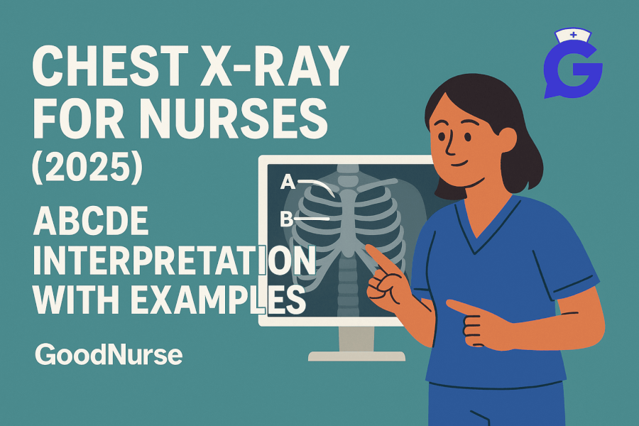
Most chest X-rays look busy until you apply a simple, repeatable checklist. This guide gives you a bedside-friendly ABCDE method, common red flags, a lines & tubes check, and mini-cases with answers—so you can choose two actions that change physiology now and two parameters that prove it worked (the exact NGN habit we teach at GoodNurse).
For perfusion/oxygenation context, keep ABG Interpretation Made Simple (2026) open. Tie radiographic findings to biomarkers with Cardiac Markers (2026): Troponin & BNP and to gas exchange with Lactate in Sepsis (2026).
Table of Contents
- The ABCDE Method (Floor Version)
- Quality Check Before You Read
- Lines & Tubes: Fast Placement Audit
- Common Patterns: What You’ll Actually See
- Red Flags & First Actions
- Mini Practice Cases (with Answers)
- FAQs
- Further Reading
🎯 Free NCLEX quiz!
Test your knowledge - new quizzes added weekly!
The ABCDE Method (Floor Version)
Use the same order every time. It’s fast, NGN-friendly, and works even when the film quality isn’t perfect.
A — Airway & Mediastinum
- Trachea midline? Any deviation (tension pneumothorax, large effusion, mass)?
- Carina visible? Mainstem intubation risk if ETT is too deep.
B — Breathing (Lungs & Pleura)
- Compare right/left lung fields for asymmetry, interstitial markings, consolidation, atelectasis, pneumothorax.
- Look for pleural lines, costophrenic angle blunting (effusions).
C — Circulation (Cardiac silhouette & vessels)
- Cardiac size (rough guide: cardiothoracic ratio on PA view).
- Pulmonary vasculature: cephalization, Kerley B lines, perihilar “bat-wing” edema patterns.
D — Diaphragm & Below
- Diaphragm domes, sharp costophrenic angles, free air under diaphragm (perforation).
- Gastric bubble on the left; herniation vs elevated hemidiaphragm.
E — Everything else (Bones, soft tissues, devices)
- Ribs, clavicles, scapulae for fractures or lytic lesions.
- Soft-tissue emphysema, subcutaneous air; device location (lines, tubes, pacers).
Professor’s note: If the client is unstable, treat physiology first (oxygenation, perfusion) and use the X-ray to confirm your suspicions—not to delay care.
🥇Voted #1 Nursing Study Tool.
Personalized AI Tutor + Instant Answers to All Your Questions. 100% Money Back Guarantee!
Quality Check Before You Read
- Projection: PA vs AP (AP enlarges the heart—don’t over-call cardiomegaly).
- Rotation: Medial clavicle heads equidistant from spinous processes.
- Inspiration: ~10 posterior ribs on a good inspiratory film.
- Penetration: Spine just visible through the heart—not too dark, not too light.
- Timing: Compare to previous film and clinical status (are we getting better or worse?).
Lines & Tubes: Fast Placement Audit
- Endotracheal tube (ETT): Tip ~3–5 cm above carina (varies with neck flexion/extension).
- Nasogastric/OG tube: Tip in stomach beyond the diaphragm; no coiling in esophagus.
- Central line: Tip in lower SVC near cavoatrial junction (not in the right atrium).
- Chest tube: Side holes should be within the thoracic cavity.
- Pacemaker/ICD: Leads coursing to RA/RV appropriately.
- A-line (radial/femoral) won’t be on CXR but chest lines often accompany a-line care—pair with Arterial Lines & MAP for accuracy checks.
Cross-training references: ABG Interpretation (2026) for gas exchange, and Coagulation Studies (2026) when procedures or bleeding risk are in play.
Common Patterns: What You’ll Actually See
1) Lobar consolidation (pneumonia)
- Clues: Dense opacity with air bronchograms; often lobar boundaries.
- Actions: Oxygen to target; support work of breathing; expect cultures/antibiotics per order.
- Parameters: SpO₂, RR/WOB, temperature, symptom trajectory.
- Practice links: NGN Med-Surg Cases for integrated scenarios.
2) Pulmonary edema (cardiogenic)
- Clues: Perihilar “bat-wing” opacities, Kerley B lines, pleural effusions, enlarged heart (PA view).
- Actions: Oxygen; position upright; anticipate diuresis/afterload reduction per orders; monitor perfusion.
- Parameters: SpO₂, MAP, UOP, dyspnea relief.
- Tie to biomarkers: Cardiac Markers.
3) Atelectasis
- Clues: Volume loss, tracheal deviation toward the lesion, diaphragmatic elevation, linear “plate-like” opacities.
- Actions: Incentive spirometry, mobilization, pain control, suctioning as ordered.
- Parameters: SpO₂, RR/WOB, auscultation improvement.
4) Pleural effusion
- Clues: Meniscus sign, blunted costophrenic angles; large effusions can shift mediastinum.
- Actions: Oxygen to target; elevate HOB; anticipate drainage/diuresis per plan; monitor hemodynamics.
- Parameters: SpO₂, MAP, dyspnea, symptom relief.
5) Pneumothorax (PTX)
- Clues: Visible pleural line with no markings beyond; tension PTX may shift trachea away and flatten hemidiaphragm.
- Actions: If unstable, prepare for emergent decompression per algorithm; oxygen; monitor closely.
- Parameters: SpO₂, RR/WOB, MAP, symptom relief.
6) COPD hyperinflation
- Clues: Flattened diaphragms, increased retrosternal airspace, barrel chest appearance.
- Actions: Oxygen to target; bronchodilator and steroid protocols per orders; watch for air trapping/fatigue.
- Parameters: SpO₂, RR/WOB, ABG trajectory.
7) Lines/tubes malposition
- Clues: ETT mainstem, NG coiled, central line too high/low.
- Actions: Notify and prepare for repositioning; keep client safe (oxygenation first).
- Parameters: SpO₂, breath sounds symmetry, post-adjustment film if ordered.
Red Flags & First Actions
- Tracheal deviation + hypotension + unilateral lucency → treat as tension PTX risk: oxygen, rapid escalation for decompression.
- Massive white-out hemithorax → think large effusion or mainstem intubation with atelectasis; assess breath sounds and tube depth.
- Device malposition (ETT, NG, central line) → correct placement promptly to avoid harm.
- Sudden airspace changes with fever → pneumonia vs aspiration; support oxygenation and follow diagnostic pathway.
For linking radiology to labs and decisions, keep CBC & CMP (2026) handy, and ensure rhythm/hemodynamics are stable with EKG Basics (2026).
Mini Practice Cases (with Answers)
Case 1 — The right mainstem
- Stem: Post-intubation CXR: ETT ~1 cm above carina; right lung hyperinflated; left lung looks atelectatic; SpO₂ drifting down.
- Question: Best action pair?
- Answer: Withdraw ETT slightly per order, re-assess breath sounds and SpO₂; repeat film if ordered.
- Rationale: Mainstem intubation—oxygenation risk. Parameters: SpO₂, RR/WOB, symmetric breath sounds.
Case 2 — New unilateral opacity post-op
- Stem: Day 1 post-op; linear basilar opacities and mild elevation of right hemidiaphragm.
- Question: Priority?
- Answer: Incentive spirometry, mobilization, analgesia optimization.
- Rationale: Post-op atelectasis. Parameters: SpO₂, RR/WOB, auscultation.
Case 3 — Bat-wing pattern with orthopnea
- Stem: Dyspnea, orthopnea; perihilar opacities and small effusions; mild cardiomegaly on PA film.
- Question: First actions?
- Answer: Oxygen, upright positioning; anticipate diuresis per orders; monitor UOP and MAP.
- Rationale: Likely cardiogenic edema. Parameters: SpO₂, MAP, UOP, dyspnea relief.
Case 4 — Suspected PTX after line placement
- Stem: Central line placed; client pleuritic pain; CXR shows pleural line with no distal markings.
- Question: Nursing priorities?
- Answer: Oxygen, notify provider, prepare for decompression if unstable; continuous monitoring.
- Rationale: Iatrogenic pneumothorax risk. Parameters: SpO₂, RR/WOB, MAP.
Case 5 — Large effusion with shift
- Stem: Dyspnea, dullness to percussion; white-out with mediastinal shift.
- Question: Best action?
- Answer: Oxygen, elevate HOB; anticipate drainage per plan; monitor hemodynamics.
- Rationale: Large effusion impairing ventilation. Parameters: SpO₂, MAP, symptom relief.
Professor’s note: When two answers seem reasonable, pick the one that improves oxygenation or ventilation now, then choose parameters that change within minutes (SpO₂, RR/WOB, MAP).
FAQs
Do I diagnose from an AP portable?
No—AP enlarges the heart and can distort anatomy. Use it for trend + safety checks and confirm on better imaging when feasible.
How do I avoid over-calling pneumonia?
Pair the film with fever, WBC, sputum, and auscultation; compare to prior films. Atelectasis and edema can mimic consolidation.
What proves my intervention worked?
Choose SpO₂, RR/WOB, and MAP/UOP (if perfusion issues) as immediate parameters; imaging changes lag behind.
Further Reading
Download our free printable Lab Values cheat sheet. Enter your email below and we'll send it right over.